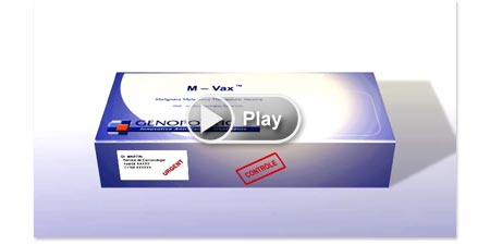
Principle of preparation and MOA of autologous anticancer vaccine
Step 1 - Extraction of a tumuor
During surgery aimed at removing or reducing the tumour charge of a patient, a tumour is removed and shipped to a laboratory in a specific refrigerated container.
Step 1 - Extraction of a tumuor
During surgery aimed at removing or reducing the tumour charge of a patient, a tumour is removed and shipped to a laboratory in a specific refrigerated container.
Principle of preparation and MOA of autologous anticancer vaccine
Step 2 - Isolation of cancer cells from the tumour
In the laboratory, the tumour is controlled and enters a well-defined production protocol, the first step of which is to isolate the tumour cells.
Step 2 - Isolation of cancer cells from the tumour
In the laboratory, the tumour is controlled and enters a well-defined production protocol, the first step of which is to isolate the tumour cells.
Principle of preparation and MOA of autologous anticancer vaccine
Step 3 - Treatment of tumour cells to render them immunogenic
The tumour cells are then modified, irradiation and haptenisation, two steps that will induce an immune response upon vaccination of the patient.
Step 3 - Treatment of tumour cells to render them immunogenic
The tumour cells are then modified, irradiation and haptenisation, two steps that will induce an immune response upon vaccination of the patient.
Principle of preparation and MOA of autologous anticancer vaccine
Step 4 - Sampling of the vaccine
Treated cancer cells are sampled in 8 vaccine units placed into a container labelled with the patient name (autologous and individualised treatment).
Step 4 - Sampling of the vaccine
Treated cancer cells are sampled in 8 vaccine units placed into a container labelled with the patient name (autologous and individualised treatment).
Principle of preparation and MOA of autologous anticancer vaccine
Step 5 - Vaccine injection
The anti-cancer autologous vaccine is injected intradermally under medical control according to a precise 7 weeks time schedule followed by a last injection after 6 months.
Step 5 - Vaccine injection
The anti-cancer autologous vaccine is injected intradermally under medical control according to a precise 7 weeks time schedule followed by a last injection after 6 months.
Principle of preparation and MOA of autologous anticancer vaccine
Step 6 - Principle of recognition of the modified tumour cells by the immune system
The aim of the anti-cancer vaccine is to induce an individualised immune response against its very own tumour antigens, which will block or delay reappearance of tumour. This response is due to the chemical modification of tumour cells from the vaccine that will break the tolerance against the tumour antigens.
Principle of preparation and MOA of autologous anticancer vaccine
Step 7 - Recognition of cancer cells by the immune system
The induced immune response will propagate to the tumours via the immune system. Breakdown of the tolerance to tumour antigens involves specific T-lymphocytes that lead to an inflammatory response. This response would be due to the infiltration of the tumour by CD8+ T-lymphocytes, in situ production of γ interferon and clonal expansion of T-lymphocytes.


